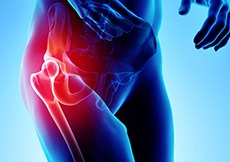
Hip Anatomy
The hip joint is the largest weight-bearing joint in the human body. It is also referred to as a ball and socket joint and is surrounded by muscles, ligaments and tendons. The thighbone or femur and the pelvis join to form the hip joint.
Any injury or disease of the hip will adversely affect the joint's range of motion and ability to bear weight.
The hip joint is made up of the following:
- Bones and joints
- Ligaments of the joint capsule
- Muscles and tendons
- Nerves and blood vessels that supply the bones and muscles of the hip
Bones and Joints of the Hip
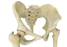
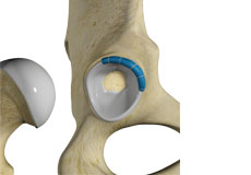
The hip joint is the junction where the hip joins the leg to the trunk of the body. It is comprised of two bones: the thighbone or femur, and the pelvis, which is made up of three bones called ilium, ischium and pubis.
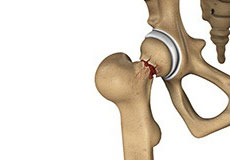
The ball of the hip joint is made by the femoral head while the socket is formed by the acetabulum. The acetabulum is a deep, circular socket formed on the outer edge of the pelvis by the union of three bones: ilium, ischium and pubis. The lower part of the ilium is attached by the pubis while the ischium is considerably behind the pubis. The stability of the hip is provided by the joint capsule or acetabulum and the muscles and ligaments that surround and support the hip joint.
The head of the femur rotates and glides within the acetabulum. A fibrocartilaginous lining called the labrum is attached to the acetabulum and further increases the depth of the socket.
The femur is one of the longest bones in the human body. The upper part of the thighbone consists of the femoral head, femoral neck, and greater and lesser trochanters. The head of the femur joins the pelvis (acetabulum) to form the hip joint. Next to the femoral neck, there are two protrusions known as greater and lesser trochanters which serve as sites of muscle attachment.
Articular cartilage is the thin, tough, flexible and slippery surface lubricated by synovial fluid that covers the weight-bearing bones of the body. It enables smooth movements of the bones and reduces friction.
Ligaments of the Hip Joint
Ligaments are fibrous structures that connect bones to other bones. The hip joint is encircled with ligaments to provide stability to the hip by forming a dense and fibrous structure around the joint capsule. The ligaments adjoining the hip joint include:
- Iliofemoral ligament:his is a Y-shaped ligament that connects the pelvis to the femoral head at the front of the joint. It helps in limiting over-extension of the hip.
- Pubofemoral ligament:his is a triangular shaped ligament that extends between the upper portion of the pubis and the iliofemoral ligament. It attaches the pubis to the femoral head.
- Ischiofemoral ligament: his is a group of strong fibers that arise from the ischium behind the acetabulum and merge with the fibers of the joint capsule.
- Ligamentum teres:his is a small ligament that extends from the tip of the femoral head to the acetabulum. Although it has no role in hip movement, it does have a small artery within that supplies blood to a part of the femoral head.
- Acetabular labrum: he labrum is a fibrous cartilage ring which lines the acetabular socket. It deepens the cavity increasing the stability and strength of the hip joint.
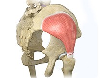
Muscles and Tendons of the Hip Joint
A long tendon called the iliotibial band runs along the femur from the hip to the knee and serves as an attachment site for several hip muscles including the following:
- Gluteal:hese are the muscles that form the buttocks. There are three muscles (gluteus minimus, gluteus maximus, and gluteus medius) that attach to the back of the pelvis and insert into the greater trochanter of the femur.
- Adductors:hese muscles are in the thighs which help in adduction, the action of pulling the leg back towards the midline.
- Iliopsoas:his muscle is in front of the hip joint and provides flexion. It is a deep muscle that originates from the lower back and pelvis, and extends up to the inside surface of the upper part of the femur.
- Rectus femoris:his is the largest band of muscles located in front of the thigh. They are also called hip flexors.
- Hamstring muscles:hese begin at the bottom of the pelvis and run down the back of the thigh. Because they cross the back of the hip joint, they help in extension of the hip by pulling it backwards.
Nerves and Arteries of the Hip Joint
Nerves of the hip transfer signals from the brain to the muscles to aid in hip movement. They also carry the sensory signals such as touch, pain, and temperature back to the brain.
The main nerves in the hip region include the femoral nerve in the front of the femur and the sciatic nerve at the back. The hip is also supplied by a smaller nerve known as the obturator nerve.
In addition to these nerves, there are blood vessels that supply blood to the lower limbs. The femoral artery, one of the largest arteries in the body, arises deep in the pelvis and can be felt in front of the upper thigh.
Hip Movements
All the anatomical parts of the hip work together to enable various movements. Hip movements include flexion, extension, abduction, adduction, circumduction, and hip rotation.
Conditions

Osteoarthritis of the Hip
Osteoarthritis, also called degenerative joint disease, is the most common form of arthritis. It occurs most often in the elderly.
Inflammatory Arthritis of the Hip
The inflammation of the joints is referred to as arthritis. Inflammation arises when the smooth lining called cartilage at the ends of bones wears away.
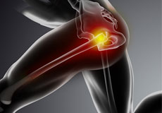
Hip Injury
The hip joint is one of the most important and flexible joints in the human body which allows us to walk, run, bend and perform physical activities.
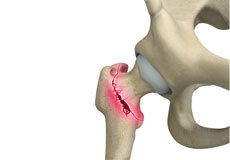
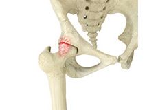
Hip Fracture
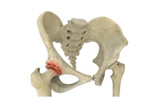
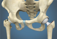
The hip joint is a “ball and socket” joint. The “ball” is the head of the femur or thighbone, and the “socket” is the cup-shaped acetabulum.
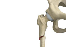
Femur Fracture
The femur or thigh bone is the longest and strongest bone in the body, connecting the hip to the knee. A femur fracture is a break in the femur.
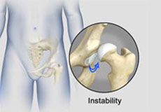
Hip Instability
The hip plays an important role in supporting your upper body weight while standing, walking and running, and hip stability is crucial for these functions.
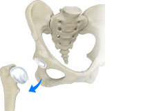
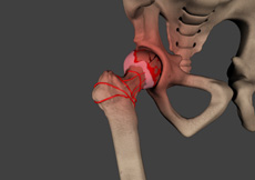
Hip Dislocation
The hip joint is a “ball and socket” joint. The “ball” is the head of the femur or thighbone, and the “socket” is the cup-shaped acetabulum.
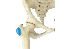
Hip Bursitis
Hip bursitis is a painful condition caused by the inflammation of a bursa in the hip.
Hip Ligament Injuries
Injuries to the hip ligaments are commonly called a hip sprain and can range from minor tears of the ligaments to more serious injuries involving the hip muscles, tendons or bone.
Hip Abductor Tears
Hip abductors are a major group of muscles found in the buttocks. It includes the gluteus maximus, gluteus medius, gluteus minimus, and tensor fascia lata muscles.
Gluteus Tendon Tear
The gluteal muscles (situated in the buttocks) are necessary for the stability and movement of the hip joints.
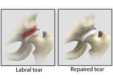
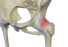
Hip Labral Tear
The hip joint is a ball and socket joint in which the head of the femur is the ball and the acetabulum forms the socket. The labrum helps to deepen the socket and provide stability to the joint.
Femoroacetabular Impingement
Femoroacetabular impingement (FAI) is a condition characterized by excessive friction in the hip joint from the presence of bony irregularities.
Avascular Necrosis
Avascular necrosis, also called osteonecrosis, is a condition in which bone death occurs because of inadequate blood supply. Lack of blood flow may occur when there is a fracture in the bone or a joint dislocation that may damage nearby blood vessels.
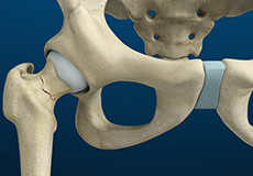
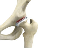
Stress Fracture of hip
Stress fractures of the hip are a break in the upper part of the thigh bone (femur) that fits into the socket of the hip joint. It can occur in any part of the hip, however, it mostly occurs just below the ball of the ball-and-socket hip joint called the femoral neck.
Subtrochanteric hip Fracture
A hip fracture is a break that occurs near the hip in the upper part of the femur or thighbone. The thighbone has two bony processes on the upper part - the greater and lesser trochanters. The lesser trochanter projects from the base of the femoral neck on the back of the thighbone.
Periprosthetic Hip Fracture
Hip replacement is a surgical procedure in which the damaged cartilage and bone are removed from the hip joint and replaced with artificial components. Any resulting fractures or breaks in the bone around the implant are called periprosthetic hip fractures.
Procedures
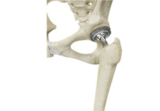
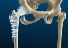
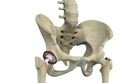
Total Hip Replacement
Total hip replacement is a surgical procedure in which the damaged cartilage and bone are removed from the hip joint and replaced with artificial components.
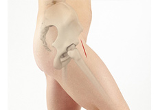
Direct Anterior Hip Replacement
Direct anterior hip replacement is a minimally invasive hip surgery to replace the hip joint without cutting through any muscles or tendons as against traditional hip replacement
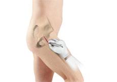
Minimally Invasive Surgery
Minimally invasive total hip replacement is a surgical procedure performed through one or two small incisions rather than the single long incision of 10–12-inches as in the traditional approach.
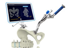
Mako Robotic Arm Assisted Hip Replacement
Robotic assisted hip replacement is an Image-guided procedure that involves the use of a specialized robotic system to assist your surgeon in performing a total hip replacement
Hip Fracture Surgery
Hip fractures involve a break that occurs near the hip in the upper part of the femur or thigh bone. The thigh bone has two bony processes on the upper part - the greater and lesser trochanters.
Total Hip Revision Surgery
During total hip replacement, the damaged cartilage and bone are removed from the hip joint and replaced with artificial components.

Outpatient Hip Replacement
Hip replacement surgery is one of the most common orthopedic surgeries performed. It involves the replacement of the damaged hip bone
Robotic Assisted Hip Replacement
Robotic assisted hip surgery is an Image-guided procedure that involves the use of a specialized robotic system to assist your surgeon in performing a total hip replacement
Hip Hemiarthroplasty
Hip hemiarthroplasty is a surgical technique employed to treat hip fractures. In this procedure, only one half (ball section) of the hip joint is substituted by a metal prosthesis.
Hip Trauma Reconstruction
Hip trauma is an injury in the hip due to the impact caused by incidents such as a car accident or a hard fall. The injury can be a bone break or dislocation or both.

Activities After Hip Replacement
Hip replacement is a surgery performed to replace parts of a diseased hip joint with a prosthesis. The goal of hip replacement is to eliminate pain
Physical Therapy for Hip
Physical therapy is an exercise program that helps you to improve movement, relieve pain, encourage blood flow for faster healing, and restore your physical function and fitness level.