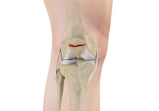
Knee Anatomy
The knee is a complex joint made up of different structures including bones, tendons, ligaments and muscles. They all work together to maintain normal function and provide stability to the knee during movement.
Having a well-functioning healthy knee is essential for our mobility and ability to participate in various activities. Understanding the anatomy of the knee enhances your ability to discuss and choose the right treatment procedure for knee problems with your doctor.
Bones of the Knee
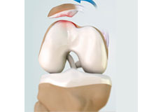
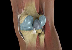
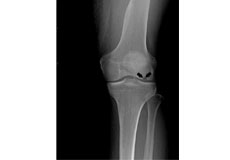
The knee is a hinge joint made up of two bones, the thighbone (femur) and the shinbone (tibia). There are two round knobs at the end of the femur called femoral condyles which articulate with the flat surface of the tibia called the tibial plateau. The tibia plateau on the inside of the leg is called the medial tibial plateau, and on the outside of the leg it is called the lateral tibial plateau.
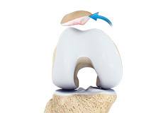
The two femoral condyles form a groove on the front (anterior) side of the knee called the patellofemoral groove. A small bone called the patella sits in this groove and forms the kneecap. It acts as a shield and protects the knee joint from direct trauma.
A fourth bone called the fibula is the other bone of the lower leg. This forms a small joint with the tibia. This joint has very little movement and is not considered a part of the main joint of the knee.
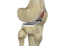
Articular Cartilage and Menisci of the Knee
Movement of the bones causes friction between the articulating surfaces. To reduce this friction, all articulating surfaces involved in movement are covered with a white, shiny, slippery layer called articular cartilage. The articulating surface of the femoral condyles, tibial plateaus and the back of the patella are covered with this cartilage. The cartilage provides a smooth surface that facilitates easy movement.
To further reduce friction between the articulating surfaces of the bones, the knee joint is lined by a synovial membrane which produces a thick clear fluid called synovial fluid. This fluid lubricates and nourishes the cartilage and bones inside the joint capsule.
Within the knee joint, between the femur and tibia, there are two C shaped cartilaginous structures called menisci. Menisci function to provide stability to the knee by spreading the weight of the upper body across the whole surface of the tibial plateau. The menisci help in load- bearing by preventing the weight from concentrating onto a small area, which could damage the articular cartilage. The menisci also act as a cushion between the femur and tibia by absorbing the shock produced by activities such as walking, running and jumping.
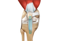
Ligaments of the Knee
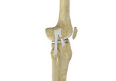
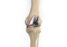
Ligaments are tough bands of tissue that connect one bone to another bone. The ligaments of the knee function to stabilize the knee joint. There are two important groups of ligaments that hold the bones of the knee joint together, collateral ligaments and the cruciate ligament.
Collateral ligaments are present on either side of the knee. They function to prevent the knee from moving too far during side to side motion. The collateral ligament on the inside is called the medial collateral ligament (MCL) and the collateral ligament on the outside is called the lateral collateral ligament (LCL).
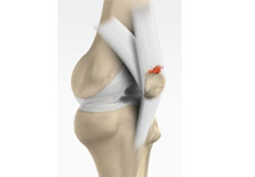
Cruciate ligaments, present inside the knee joint, control the back-and-forth motion of the knee. The cruciate ligament in the front of the knee is called anterior cruciate ligament or ACL and the cruciate ligament in the back of the knee is called posterior cruciate ligament or PCL.
Muscles of the Knee
Muscles: There are two major muscles, the quadriceps and the hamstrings, which enable movement of the knee joint. The quadriceps muscles are in the front of the thigh. When the quadriceps muscles contract, the knee straightens. The hamstrings are in the back of the thigh. When the hamstring muscles contract, the knee bends.
Tendons of the Knee
Tendons are structures that attach muscles to the bone. The quadriceps muscles of the knee meet just above the patella and attach to it through a tendon called the quadriceps tendon. The patella further attaches to the tibia through a tendon called the patella tendon. The quadriceps muscle, quadriceps tendon and patellar tendon all work together to straighten the knee. Similarly, the hamstring muscles at the back of the leg are attached to the knee joint with the hamstring tendon.
Conditions
Knee Arthritis
The joint surface is covered by a smooth articular surface that allows pain-free movement in the joint. Arthritis is a general term covering numerous conditions where the joint surface or cartilage wears out.
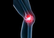
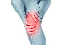
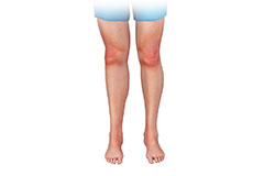
Knee Pain
Knee pain is a common condition affecting individuals of various age groups. It not only affects movement but also impacts your quality of life.
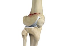
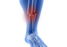
Knee Fracture
A knee fracture is a broken bone or a crack in or around the joint of the knee. This can involve the tibia (shin bone), the kneecap (patella), or femur (thighbone) where they connect with the knee.
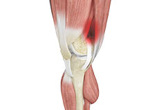
Knee Injury
Pain, swelling, and stiffness are the common symptoms of any damage or injury to the knee. If care is not taken during the initial phases of injury, it may lead to joint damage, which may end up destroying your knee.
Fractures of the Tibia
The lower leg is made up of two long bones called the tibia and fibula that extend between the knee and ankle. The tibia or shinbone is the larger of the two bones.
Fractures of the Patella
The patella or kneecap is a small bone present in the front of your knee where the thigh bone meets the shinbone. It provides protection to your knee and attachment to muscles in the front of the thigh.
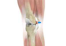
Patellofemoral Instability
Patellofemoral instability means that the patella (kneecap) moves out of its normal pattern of alignment. This malalignment can damage the underlying soft structures such as muscles and ligaments that hold the knee in place.
Knee Sprain
Knee sprain is a common injury that occurs from overstretching of the ligaments that support the knee joint. A knee sprain occurs when the knee ligaments are twisted
MCL Sprains
The medial collateral ligament (MCL), a band of tissue present on the inside of your knee joint, connects your thighbone and shinbone (bone of your lower leg).
Kneecap Bursitis
A bursa is a small fluid-filled sac found between soft tissues and bones. It lubricates and acts as a cushion to decrease friction between bones when they move.
Anterior Knee Pain
Anterior knee pain is characterized by chronic pain over the front and center of the knee joint. It is common in athletes, active adolescents (especially girls) and overweight individuals.
Knee Osteoarthritis
Osteoarthritis also called degenerative joint disease, is the most common form of arthritis. It occurs most often in older people.
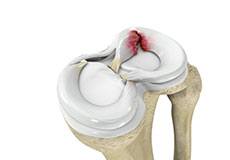
Meniscal Tears
A meniscal tear is a common knee injury in athletes, especially those involved in contact sports. A sudden bend or twist in your knee causes the meniscus to tear.
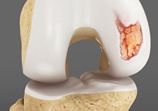
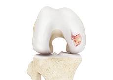
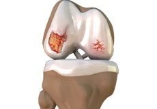
Articular Cartilage Injury
Articular or hyaline cartilage is the tissue lining the surface of the two bones in the knee joint. Cartilage helps the bones move smoothly against each other
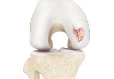
Chondral or Articular Cartilage Defects
The articular or hyaline cartilage is the tissue lining the surface of the two bones in the knee joint. Cartilage helps the bones move smoothly against each other and can withstand the weight of your body
Quadriceps Tendon Rupture
The quadriceps tendon is a thick tissue located at the top of the kneecap. It works together with the quadriceps muscles to allow us to straighten our leg.
Patellar Tendon Rupture
The patellar tendon works together with the quadriceps muscle and the quadriceps tendon to allow your knee to straighten out.
Chondromalacia Patella
The patella, also called the kneecap, is a small bone present on the front of your knee joint. The underside of the patella is covered by cartilage that allows smooth gliding of the knee with movement.
Baker’s Cyst
Baker’s cyst, in some cases, does not cause any pain and may go unnoticed. However, you may experience symptoms such as swelling behind your knee and legs, stiffness behind the knees, slight pain in the knee towards the upper calf (especially when you bend your knee or straighten it completely).
Iliotibial Band Syndrome
An iliotibial band is a tough group of fibers that runs from the iliac crest of the hip along the outside of the thigh, till the outer side of the shinbone, just below the knee joint. Its function is to coordinate with the thigh muscles and provide stability to the knee joint.
Unstable Knee
Damage to any of these supportive structures causes instability of the knee joint. An unstable knee can be caused by the sudden twisting of the knee, tears of the meniscus, ligament or capsule, osteoarthritis of the knee (wear and tear of the cushioning cartilage tissue between the bones) and sports injuries.
Osteonecrosis of the Knee
Osteonecrosis is a condition in which the death of a section of bone occurs because of lack of blood supply to it. It is one of the most common causes of knee pain in older women. Women over 60 years of age are commonly affected, three times more often than men.
Periprosthetic Knee Fractures
Knee replacement, also called knee arthroplasty, is a surgical procedure in which the worn-out or damaged surfaces of the knee joint are removed and replaced with artificial implants. Any resulting fractures or breaks in the bone around the implant are called periprosthetic knee fractures.
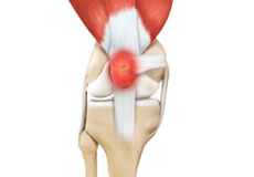
Ligament Injuries
Knee problems may arise if any of these structures get injured by overuse or suddenly during sports activities. Pain, swelling, and stiffness are the common symptoms of any damage or injury to the knee.
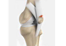
Patellar Dislocation/Patellofemoral Dislocation
Patellar dislocation occurs when the patella moves out of the patellofemoral groove, (trochlea) onto the bony head of the femur. If the kneecap partially comes out of the groove, it is called subluxation; if the kneecap completely comes out, it is called dislocation (luxation).
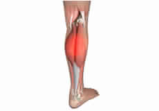
Medial Gastrocnemius Strain
A medial gastrocnemius strain (MGS), also sometimes called “tennis leg”, is an injury to the calf muscle in the back of the leg. It occurs when the calf muscle is stretched too far resulting in a partial or total tear or rupture within the muscle.
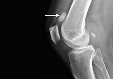
Loose Bodies in the Knee
Loose bodies are fragments of detached cartilage or bone inside the knee joint. These fragments may be free floating (unstable) or may be trapped (stable) within the joint.
Procedures
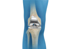
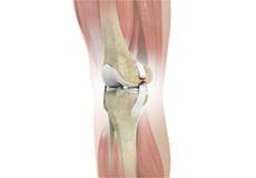
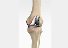
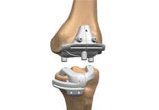
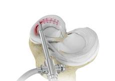
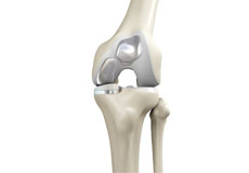
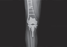
Total Knee Replacement
Total knee replacement, also called total knee arthroplasty, is a surgical procedure in which the worn out or damaged surfaces of the knee joint are removed and replaced with an artificial prosthesis
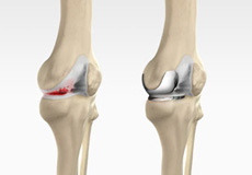
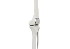
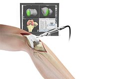
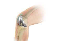
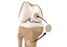
Robotic Unicondylar Knee Replacement
A unicondylar knee replacement is a procedure to replace part of the knee joint with a prosthetic implant to relieve pain and improve the function of the joint. Advances in technology have allowed this procedure to be performed in a minimally invasive manner with robotic assistance.
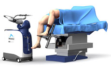
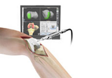
Mako Robotic Arm Assisted Total Knee Replacement
Mako Robotic-Arm Assisted Total Knee Replacement is a treatment option for adults living with mid to late-stage osteoarthritis (OA) of the knee.
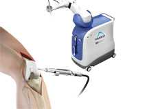
Mako Robotic Arm Assisted
Mako Robotic-Arm Assisted Technology provides you with a personalized surgical plan based on your unique anatomy. First, a CT scan of the diseased hip or knee joint is taken.
Revision Knee Replacement
Revision knee replacement surgery involves replacing a part or all your previous knee prosthesis with a new prosthesis. Although total knee replacement surgery is successful
Visionaire Patient-Matched Technology for Total Knee Replacement
Visionaire is an advanced surgical technology for a joint replacement that is based on the patient’s specific anatomy.
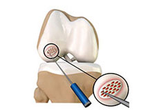
Knee Arthroscopy
Knee arthroscopy is a common surgical procedure performed using an arthroscope, a viewing instrument, to diagnose or treat a knee problem.
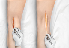
Minimally Invasive Knee Joint Replacement
Total knee replacement is a very successful surgical treatment for knee arthritis. Over the years, minimally invasive knee replacement surgical techniques have been developed
Patellofemoral Knee Replacement
Traditionally, arthritis in only one compartment of the knee is treated by partial knee replacement surgery. Patellofemoral knee replacement is a minimally invasive surgical option
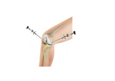
Computer Navigation for Total Knee Replacement
Computer navigation provides your surgeon with real-time 3-D images of your mapped knee and the surgical instruments during surgery.
Outpatient Total Knee Replacement
Total knee replacement is the surgical treatment for knee arthritis, where the damaged knee is removed and replaced with an artificial knee implant.
Robotic Assisted Knee Replacement
Robotic-assisted knee replacement surgery is an alternative to the conventional knee replacement procedure.

After Knee Replacement
After knee replacement surgery, once the anesthesia wears off, you will start to experience pain, for which your doctor will prescribe medication.
Quadriceps Tendon Repair
Quadriceps tendon is a thick tissue located at the top of the kneecap. The quadriceps tendon works together with the quadriceps muscles to allow us to straighten our leg.
Knee Fracture Surgery
A knee fracture is a broken bone or a crack in or around the joint of the knee. This can involve the tibia (shin bone), the kneecap (patella), or femur (thighbone) where they connect with the knee.
Meniscal Surgery
Meniscal surgery is a surgical procedure employed for the treatment of torn or damaged meniscal tissues in the knee. It is mostly performed as a minimally invasive keyhole procedure.
Arthroscopic Debridement
Osteoarthritis is a most common form of arthritis which affects the articular cartilage (tissue covering the ends of the bones) of the knee and also other joints such as shoulder, hip, ankle, and foot.
Bicompartmental Knee Resurfacing
Bicompartmental knee resurfacing is a less invasive surgical alternative to total knee replacement surgery, where instead of all the compartments being replaced only 2 of the 3 compartments
Chondroplasty
Chondroplasty is a surgical procedure to repair and reshape damaged cartilage in a joint. The procedure involves smoothing degenerative cartilage and trimming any unstable flaps of cartilage.
Non-Surgical Knee Treatments
The knee is a complex joint which consists of bone, cartilage, ligaments, and tendons that make joint movements easy and at the same time it is more susceptible to various kinds of injuries.
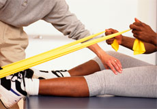
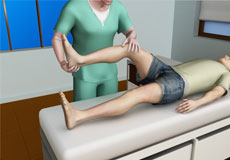
Physical Therapy for Knee
Physical therapy is an exercise program that helps you to improve movement, relieve pain, encourage blood flow for faster healing, and restore your physical function and fitness level.
Periprosthetic Knee Fracture Fixation
Periprosthetic knee fracture fixation is a procedure performed to stabilize a fracture that occurs in the bone present around a knee prosthesis. The fracture may involve the lower part of the thighbone (femur), the kneecap (patella) or the upper part of the shinbone (tibia).
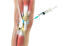
Viscosupplementation
Viscosupplementation refers to the injection of a hyaluronan preparation into the joint. Hyaluronan is a natural substance present in the joint fluid that assists in lubrication. It allows the smooth movement of the cartilage-covered articulating surfaces of the joint.